Celebrating 50 Years of Advancing University of Iowa Research
The University of Iowa's Central Microscopy Research Facility provides a wide variety of microscopy techniques for biomedical investigators, an experienced staff, and support for the beginner and experienced investigator. As one of the best equipped university core microscopy facilities in the nation, our microscopy team is ready to fully support your research program.
-------------------- CMRF News -------------------
NEW CMRF Reservation App: BookIT
The CMRF has changed to BookIT scheduling and billing app. Calpendo is no longer operational. Please use BookIT moving forward. The link to the app is:
BookitLab
If you have any questions please contact a CMRF staff member.
The results are in for the 2024 Art in Science Contest!!
The winners are:
-
1st place, Wen Wen
-
2nd place, Shubha Murthy
-
3rd place, Israel Wipf
-
4th place tie, Milad Arzani and Yan Zhang
Click on a name to see their winning image.
Congratulations to the winners! Prizes this year included binoculars from Zeiss, gift bags and $50 Java House gift certificates from Thomas/Leica and Stainless travel mugs from Nikon. Also, their their images will be featured in the 2025 CMRF Calendar.
And thank you to all who submitted images this year. We can't wait to see what images you submit next year!
The goal of The Iowa Art in Science Contest is to recognize the combination of outstanding scientific discovery and artistic appeal inherent to microscopy research. Images from any mode of microscopy can be used, and any form of colorization, manipulation and compositing is completely acceptable. Be creative, have fun!
Also, the 2025 CMRF calendar featuring 2024 Art in Science images is now available. Stop by the CMRF and pick one up, or contact Randy Nessler.
CMRF Hires New Research Assistant
Benjamin Elwood started as a Core Facility Research Assistant on December 1, 2024. Benjamin was a research assistant in Ophthalmology before joining the CMRF. Welcome, Ben!
New Equipment at the CMRF
The Hitachi 7800 TEM has been upgrade to include STEM, motorized Z-axis adjustment, selected area electron diffraction aperture, primary beam spot mask and a Bruker energy dispersive spectrometer. These upgrades will provide additional tools to support material and biological research.
FRAP and FRET have been Installed on the Leica SP8 Confocal/STED, The new modules use a “Wizard” format to guide you through configuration and data collection. FRAP and FRET software guides can be found on the SP8 website.
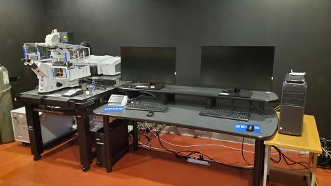
- A Zeiss LSM980 laser scanning confocal microscope with AiryScan 2 has been installed. The Airyscan 2 is a superresolution approach to imaging common fluorescent compounds. The 32-element array detector has 4-8X higher signal-to-noise over conventional confocals, with 2X higher spatial resolution in all three dimensions. The LSM980 is based on an inverted light microscope equipped with an environmental chamber for live cell imaging. Cover slipped slides can also be imaged by presenting the cover glass to the objective lens.
- A new Olympus BX63 upright light/fluorescent microscope equipped with a DP74 color CMOS camera was recently installed. A motorized stage and CellSense software allow automated tiling of slides.
- We have installed a new Tanner Scientific TN-1700 Paraffin Embedding Center that consists of a paraffin dispenser along with warm and cold plates.
- A Denton 502B thermal evaporator system was recently installed. This system allows for the evaporation of carbon and metals over samples, as well as glow discharging TEM grids.
The CMRF Announces the Purchase of a state-of-the-art Zeiss LSM 980 AiryScan 2 Confocal Microscope
The CMRF recently installed an inverted Zeiss LSM 980 AiryScan 2 confocal microscope. This next-generation confocal has improved speed, sensitivity, and resolution. The AiryScan 2 detector and Multiplex upgrade deliver fast, consistent, and quantitative Supperresolution. The system has full environmental control to support live-cell imaging experiments, while also having the capability to image standard cover-slipped slides.

Key features of the system include:
- 2 liquid-cooled MA-PMT detectors and 1 high-sensitivity 32-channel GaAsP array detector with full emission tunability
- AiryScan 2 superresolution detector, enabling 120 nm resolution in XY, 350 nm in Z, works with all available lasers, and doubles as an additional GaAsP confocal detector
- High-speed Z-piezo stage for fast volume imaging
- Observer 7 inverted stand optimized for long-term live-cell imaging with automated scanning stage, full environmental control (temperature, CO2, humidity) and Definite Focus 2 focus-maintaining system
- Argon and diode laser lines 405, 445, 488, 514, 561, 594 and 639 nm
- ZEN 3.2 software package including modules for Z-stack, time-lapse, photobleaching/photoactivation, spectral unmixing, automated image analysis, FRET, colocalization, autofocus, 3D rendering, tiles and ratiometric tools
- Transmitted light detector
- Full set of objectives includes 10x/0.3, 20x/0.8, 40x/1.2 water, 40x/1.3 oil, 63x/1.4 oil
Talk to your CMRF staff contact for more information and to arrange a demo or training.
Leica SP8 Confocal-STED Super-resolution System Upgraded
775 nm Depletion Laser Added
The CMRF’s Leica Stimulated Emission Depletion (STED) super-resolution imaging system, based on an SP8 laser scanning confocal microscope, has recently been upgraded. STED delivers resolution beyond the theoretical limit of light, and fills the resolution gap between standard confocal and electron microscopy. The system now features a 775 nm pulsed depletion laser, in addition to the existing 660 nm depletion laser. This addition will simplify multiple label STED experiments, increase resolution (<40 nm), and reduce photobleaching. The STED system also now features a 93x glycerol lens that will facilitate imaging at greater depth. Follow this link for more information on the system, and sample preparation guides.
Below is an example of the dramatic increase in resolution provided by STED. The sample is 23 nm beads labeled with ATTO647N (top two images) and Alexa 594. Confocal images on the left, 775 nm STED on the right. Note the typical diffraction-limited 200 nm spots in the confocal images, and the dramatic resolution improvement in the STED images.

FRAP and FRET Wizard Installed on Leica SP8 Confocal/STED
Also, the CMRF has upgraded the Leica SP8 Confocal/STED microscope with FRAP and FRET software. The new modules use a “Wizard” format to guide you through configuration and data collection. FRAP and FRET software guides can be found on the SP8 website.
High-end Workstation for Huygen's Pro Deconvolution and Imaris 3D and 4D Real-time Interactive Visualization and Analysis Software.
To process, analyze and visualize STED and confocal data sets, the CMRF maintains current copies of SVI’s Huygen’s Pro and Oxford’s Imaris. Huygen’s is the gold standard deconvolution app, increasing resolution and removing noise from confocal and STED z-stacks. Imaris is the world’s leading 3D-4D image visualization and analysis package, providing sophisticated, easy-to-use volume, distance and tracking modules. Both apps are installed on a custom-built high-end PC workstation.
Huygen's Pro features easy-to-use wizards that guide the user through the deconvolution process. Background flare is removed, and resolution can be be improved by 2x.
With just a few steps, Imaris delivers quantitative volumetric and motion-analysis results. Custom Imaris modules are available for neuroscience, (e.g. dendrite and spine analysis,) and co-localization studies. Other available features include:
- Imaris Measurement Pro: Shape, size and intensity-based quantification.
- Imaris Track: Imaging, tracking and motion analysis of live cells and moving objects in 2D and 3D.
- Imaris Filament Tracer: Automatic detection of neurons (dendritic trees, axons and spines,) microtubules, and other filament-like structures in 2D, 3D and 4D.
- Imaris Coloc: Quantify and document co-distribution of multiple stained biological components.
- Imaris Cell: Quantitatively examine micro relationships that exist within and between cells.
- Imaris Vantage: Compare and contrast experimental groups by visualizing image data in five dimensions as uni- or multi-variate scatterplots.
New Paraffin Processor
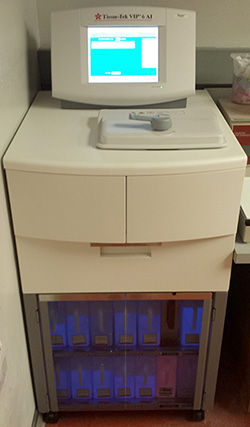
The CMRF recently installed a Tissue-Tek® VIP® 6 AI Vacuum Infiltration Paraffin Processor. This state-of-the-art system provides reproducible, high-quality processing of histological samples into paraffin. For more information, contact Chantal at chantal-allamargot@uiowa.edu.

High-Resolution Iridium Sputter Coater Available!
The CMRF has purchased a Quorum Technologies 150T ES Iridium sputter coater to support high-resolution SEM imaging. It is capable of producing coatings that are much thinner than standard Au/Pd sputter coating, which can result in higher-resolution images. The QT 150T ES is the ideal coating system for routine imaging above 50,000x. Contact CMRF staff if you are interested in using this instrument.
Office of the Vice President for Research article on the 2016 Art in Science Contest
CMRF Staff are available Monday through Friday 8am-5pm and will be happy to assist you! Call 335-8143 or email Randy Nessler for more information.


