Breadcrumb
- Home
- Instrumentation
- Light Microscopy
- MicroBrightField Stereology System
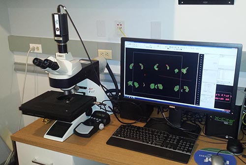
MicroBrightField Stereology System
Main navigation
- Reagents, Secondary Antibodies & Other Supplies
- Transmission Electron Microscopy
- Scanning Electron Microscopy
- Light Microscopy
- Confocal/Super Resolution Microscopy
- Sample Preparation
- Image Analysis (inst)
Eckstein Medical Research Building Room 84C

Provides accurate, unbiased Stereological estimates of the number, length, area and volume of cells or biological structures in a tissue
Instrument Specifications
- Objective lenses
- 1.5x
- 5x/0.15
- 10x/0.30
- 20x/0.50
- 40x/0.60 OIL
- Illumination Modes
- Transmitted light
- Fluorescence capabilities
- DAPI
- FITC
- Texas Red
- Other Features
- Motorized condenser
- Automatic Koehler illumination
- Motorized stage
- Z-encoder
- Software for Stereology
- Microbrightfield Stereo Investigator
- Designed based stereology
- Image analysis
- 2D anatomical mapping
- Import image data from other light microscopes(bright field, fluorescence, confocal, slide scanner) or electron microscopes
- Collect data directly from slides
- Stereology methodology
- Cavaleri method for volume measurements
- Optical fractionator for counting
- Space Balls for 3-D length measurement
- Biolucida Cloud for working with large data and sharing of files remotely
- Allows remote viewing and/or access of 2-D or 3-D virtual tissue image montages
- File support includes Jpeg2000 (.jp2, .jpf .jpx), Aperio (.svs), Nanozoomer (.ndpi, .vms, .vmu) Tiff (.tif). (Fluoview (tif), Leica and Zeiss (.lsm)
- Screen image export in .tif, .jpg or .png file formats